 english

Ostatnia modyfikacja podstrony: 12.02.2024 00:50
Retina OCT image classification
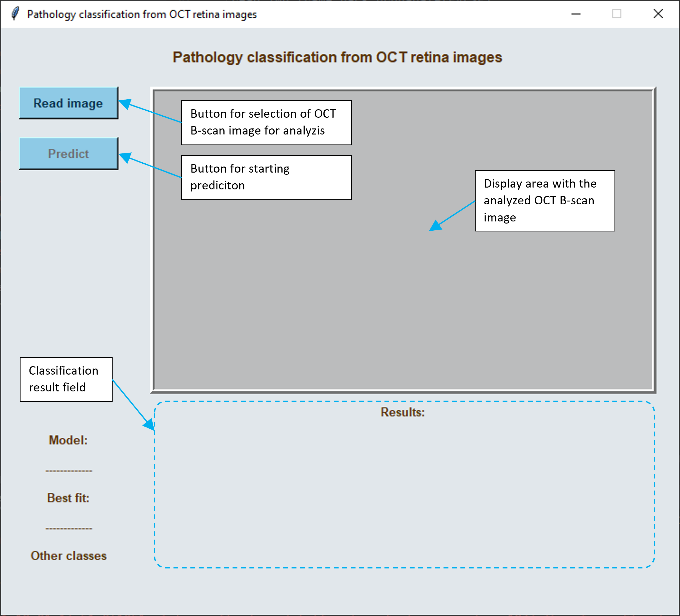
The main objective of this project is to develop an easy to use software for retina pathology classification from an OCT image by application of novel machine learning approaches with deep neural networks. The proposed classification model developed based on the idea presented here [Kermany_2018, Kaggle_Mooney] is able to classify a single OCT B-scan image as belonging to one of the following 4 classes:
- NORMAL - healthy, normative retina
- CNV - choroidal neovascularization
- DME - diabetic macular edema
- DRUSEN - present in age-related macular degeneration.
Download and usage
The Software can be downloaded from here:
- Windows version: [RePatClas_multi.zip] (780 MB)
- MacOS version: [RePatClas_dist_macOS.zip] (570 MB)
To use the Software, upack the downloaded archive and run the RePatClass file. No installation or additional libraries are required. The Software was developed on Windows 10 / MacBook Air. No additional precessing power is required (i.e. GPU card).
Important note: The tested image file name and its path cannot contain special characters (e.g. "ę", "ś", "ö")
Important note: Depending on the machine capabilities (processor and RAM) it may take up to 60 seconds to load the necessary libraries at the application startup.
For training and testing models the data from public LOCT dataset (link) was used:
- Training dataset (32 000 images): [link] (2 GB)
- Testing dataset (1 000 images): [link] (47 MB)
Scientific Representatives:
Agnieszka Stankiewicz, Tomasz Marciniak
POZNAN UNIVERSITY OF TECHNOLOGY,
Faculty of Control, Robotics and Electrical Engineering,
Institute of Automatic Control and Robotics,
Division of Signal Processing and Electronic Systems
Address: ul. Jana Pawla II 24, 61-138 Poznan, POLAND
Terms of Use
- This software is to be used only for investigational/research purposes. The Software may not be used in the diagnosis or treatment of patients.
- Please bear in mind that this Software is a "work-in-progress", and some features may not work properly. If you wish to improve this software please feel free to contact us at agnieszka.stankiewicz[at]put.poznan.pl or tomasz.marciniak[at]put.poznan.pl. Your help will be welcome!
- This software may not be used in a manner which generates income for the User.
- This software is and open-source free-of-charge implementation of algorithms developed by the Scientific Representatives.
- User agrees that Scientific Representatives shall receive appropriate recognition in any publication or presentation resulting from the use of this Software. A list of possible citations can be found below.
Papers:
1. T. Marciniak and A. Stankiewicz, "Automated Classification of VMT Pathology from Optical Coherence Tomography B-scans," 2022 Signal Processing: Algorithms, Architectures, Arrangements, and Applications (SPA), 2022, pp. 104-109, doi: 10.23919/SPA53010.2022.9927917. [IEEEXplore]
2. Stankiewicz, A.; Marciniak, T.; Dabrowski, A.; Stopa, M.; Marciniak, E.; Obara, B. Segmentation of Preretinal Space in Optical Coherence Tomography Images Using Deep Neural Networks. Sensors 2021, 21, 7521. https://doi.org/10.3390/s21227521. [Sensors]